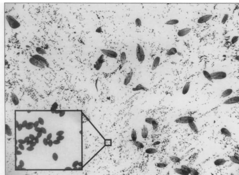
Ophryocystis elektroscirrha (O.E.) spores are much smaller than Monarch butterfly scales. The size of O.E. spores is typically in the range of 4 to 6 micrometers (µm) in length, which is about 0.005mm or 0.0002 inches, or one hundred times smaller than the butterfly’s scales.
Materials Needed
- A clear sticker or transparent tape
- Microscope slides
- Microscope
- Gloves
- Forceps or tweezers
Steps for Gathering Samples
- Put on Gloves: Always wear gloves to minimize the risk of contaminating the sample or spreading the parasite.
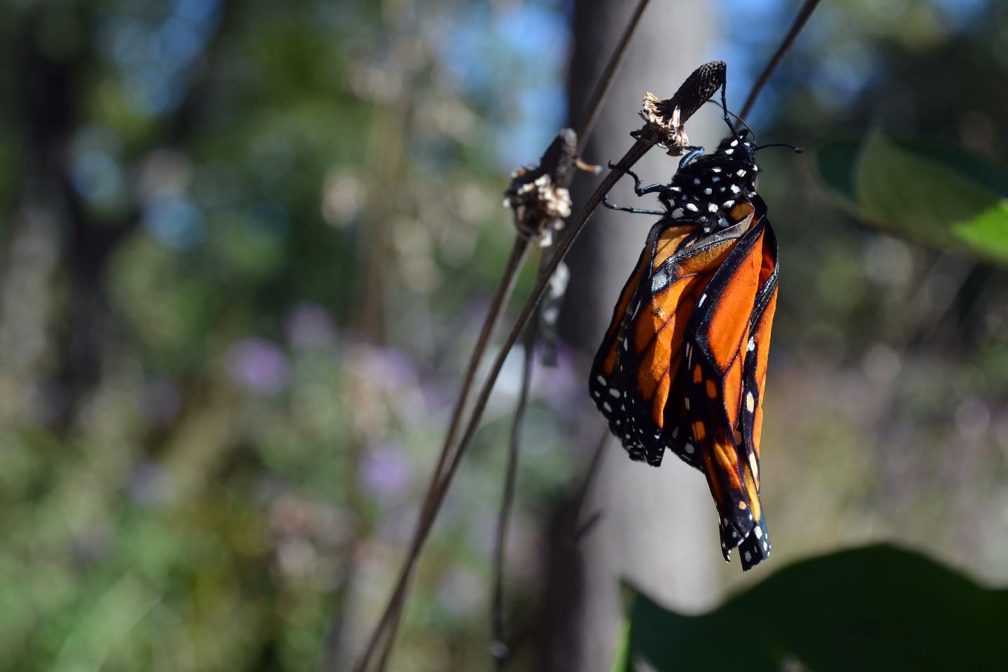
- Capture the Butterfly: Gently capture the Monarch butterfly you wish to test. Handle it carefully to avoid causing it stress or harm.
- Apply the Sticker: Gently press a clear sticker or a piece of transparent tape against the butterfly’s abdomen, ensuring you’ve made contact with the scales.
- Remove and Place on Slide: Carefully peel off the sticker and place it onto a microscope slide.
Examining Under a Microscope
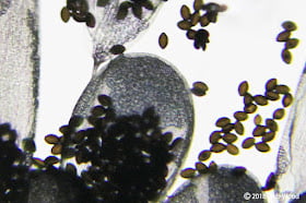
- Slide Preparation: Place the slide under the microscope, adjusting the focus until the sample is clear.
- Identification: Look for oval-shaped spores that are characteristic of O.E. They are usually quite distinct and easy to spot. There are very small, and require 400X magnification to see clearly. Butterfly scales are much larger than O.E. spores, but are also oval, so they can be mistaken for O.E. spores.
- Level of Infection: If you observe a high number of spores, the butterfly is likely heavily infected. A few spores indicate a lower level of infection.
- Record Findings: Make a note of your observations for future reference or for sharing with researchers.
Conclusion
O.E. is a significant concern for Monarch and Queen butterflies, affecting their health, longevity, and even the broader ecosystem. While researchers continue to study this parasite, citizen scientists can also play a role. By understanding how to test for O.E., we can better gauge its prevalence and potentially help in conservation efforts… one day at a time!
The Adventures of Johnny Butterflyseed – Author Signed First Edition Children’s Book
Save the monarchs!
Johnny Butterflyseed and his fairy friend, Raven Silverwing, embark on a mission to save the rapidly disappearing butterflies. They enlist the help of Queen Venus Goldwing and her kingdom of monarchs to educate and inspire kids to become butterfly farmers. At first, Johnny faces his own internal struggle with self-doubt and fear in his ability to make a difference, but then soon develops a mindset that allows him to not only get started, but also make progress one day at a time. Through challenge after challenge, Johnny learns that he is not alone in his mission and that there are many people who want to help. Together, Johnny, Raven, and Queen Venus educate thousands of children on becoming butterfly farmers.
Davis, A. K., Altizer, S., & Friedle, E. (2004). A Non-Destructive, Automated Method of Counting Spores of Ophryocystis elektroscirrha (Neogregarinorida: Ophryocystidae) in Infected Monarch Butterflies (Lepidoptera: Nymphalidae). The Florida Entomologist, 87(2), 231–234. http://www.jstor.org/stable/3496931